The pathophysiology of sickle cell disease (SCD) involves altered biophysical properties of red blood cells (RBCs) and increased cellular adhesion, which can synergistically trigger recurrent and painful vaso-occlusive events in the microcirculatory network. RBC adhesion to the endothelial wall is heterogeneous and may initiate such occlusions by disrupting the local flow thus activating platelets and promoting subsequent cell-cell interactions. Moreover, these episodic events take place within a wide range of dynamically changing shear rates at the microscale. In order to better understand the role of shear rate on this process, we quantified shear-dependent RBC adhesion to endothelial proteins fibronectin (FN) and laminin (LN) utilizing a microfluidic system that can simulate physiologically relevant shear gradients of microcirculatory blood flow at a single flow rate.
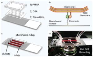
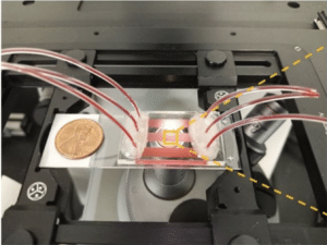
Whole blood samples were collected from 20 patients (10 males and 10 females) with homozygous SCD (HbSS). Samples were perfused through FN and LN immobilized shear-gradient microchannels (Fig. 1A) in which the shear rate continuously changes along flow direction. Computational simulations characterized the flow dynamics near the adherent RBCs (Fig. 1B). Based on the numerical results, a rectangular “field of interest (FOI)”, along which the shear rate dropped approximately three-fold, was chosen for quantification of shear-dependent RBC adhesion.
We observed changes in RBC adhesion to LN and FN in the shear gradient flow. Figure 1C and 1D show typical adhesion curves of surface adherent RBCs for an individual SCD sample within the FOI. To assess patient specific shear-dependent adhesion, we defined a parameter, “shear dependent adhesion rate (SDAR)”, which is the slope of the adhesion curves based on normalized RBC adhesion numbers.
A higher SDAR value was indicative of marked numbers of adherent RBCs that detach at higher shear rates whereas the effect of shear rate on RBC detachment was less for a lower SDAR. We observed an inverse relationship between SDAR and number of persistently adherent RBCs at high shear rates. Shear-dependent RBC adhesion to LN was heterogeneous among SCD patients. Patients with higher WBC counts constituted the low SDAR population with a threshold SDAR value of 60 (Fig. 1E, p=0.005, ANOVA). WBCs from patients with higher SDARs (and fewer persistently adhered cells) were all within the normal range. Patients in the low SDAR group also had significantly elevated absolute neutrophil counts (Fig. 1F, p=0.006, ANOVA), and ferritin levels (Fig. 1G, p=0.007, ANOVA). The mean ferritin level of those with low SDAR was nearly ten times greater than normal (mean= [3272.3 ± 791.9] μg/L vs. [784.5±219.6] μg/L). No white blood cell (WBC) adhesion was observed in the experiments.
Here, we report a novel shear dependent adhesion ratio of sickle RBCs utilizing LN and FN functionalized microchannels. The approach presented here enabled us to create a shear gradient throughout the channel which may simulate the physiological flow conditions in the post-capillary venules. We further analyzed shear-dependent RBC adhesion in a patient specific manner and identified patient groups with low and high SDAR. The findings also suggested a link between lower shear dependent sickle RBC adhesion to LN and patient clinical phenotypes including inflammation and iron overload.
Acknowledgments: This work was supported by grant #2013126 from the Doris Duke Charitable Foundation, National Heart Lung and Blood Institute R01HL133574, and National Science Foundation CAREER Award 1552782.
Figure 1: Shear-dependent sickle RBC adhesion in microscale flow. (A) Macroscopic image of the shear-gradient microchannel with the arrow indicating flow direction. (B) Velocity and shear rate contours on a 2D plane above the bottom surface. The dashed rectangular area indicates the field of interest (FOI) where the experimental data were obtained. (C, D) Typical distribution of adherent deformable and non-deformable RBCs in LN and FN functionalized microchannels with the shear gradient. Dashed lines represent the adhesion curves and the corresponding equations were used to quantify shear dependent adhesion data. Shear-dependent RBC adhesion was lower (nSDAR<60) in patients with elevated white blood cell counts (E), absolute neutrophil counts (F), and serum ferritin levels (G). The dashed rectangles indicate the normal clinical values.

