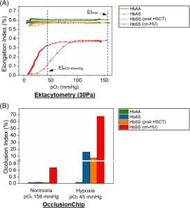
Purpose: Successful, full chimerism hematopoietic stem cell transplant (HSCT) from a matched sibling donor is curative for sickle cell disease (SCD). Current cell therapy trials of less established strategies typically have reduction in pain events as their primary endpoint, which is important, but not equivalent to a cure. Therapies with curative intent need to also be comprehensively assessed using analytical assays that provide detailed and robust functional assessment of red blood bell (RBC) properties. We hypothesize that microfluidic BioChip Assays can complement clinical assessments of efficacy in curative therapies. To test this, we assessed RBC adhesion using SCD BioChip A and RBC deformability using SCD BioChip D, before and after HSCT.
Materials and methods: Venous blood samples were collected in EDTA tubes from a patient with SCD at University Hospitals, Cleveland Medical Center. The samples were tested with the SCD Biochip A and D at multiple time points between 2016 and 2022. SCD BioChip A devices were fabricated using lamination processes and were functionalized with Intercellular Adhesion Molecule 1 (ICAM-1) and Laminin (LN). Whole blood was injected into the ICAM-1 and LN-immobilized microchannels. An inverted microscope and microscopy camera were used to obtain high-resolution images of the whole channel for processing (Adobe Photoshop, San Jose, CA) and quantification of adhered RBCs per unit area (32 mm2). Adhesion Indices for ICAM-1 and LN were calculated. SCD BioChip D devices were fabricated using standard soft lithography protocols. RBCs were isolated from whole blood, re-suspended in PBS at 20% hematocrit, and passed through the device with a constant inlet pressure. Following a wash step, the microchannel was imaged, processed and Occlusion Index (OI) was quantified.