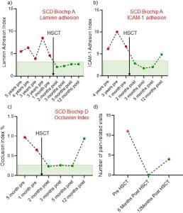
Purpose: Red blood cell (RBC) deformability is a biomarker to assess the clinical status and response to therapy in sickle cell disease (SCD). RBC deformability has been measured by ektacytometry for decades, using shear or osmolar stress. However, ektacytometry averages the RBC population, and cannot report small fractions of abnormal RBCs. A single cell-based, functional RBC deformability measurement provides essential information on the presence of unmodified cell populations that can perpetuate disease complications after the implementation of drug or cell-based therapies. There are two goals in this study: to determine the reproducibility of values generated by the standardized OcclusionChip assay and commercially available Oxygen Gradient Ektacytometry (LORRCA); and to determine if the OcclusionChip assay could complement Ektacytometry for detection of abnormal RBCs in small fractions.
Materials and methods: OcclusionChip microfluidic assay design includes a gradient of capillary network-inspired micropillar arrays embedded into the microfluidic channel forming microcapillaries from 20 μm down to 4 μm. This mimics the non-uniform, continuously changing capillaries in the capillary bed, retaining RBCs with significantly decreased deformability in the upstream array with coarser openings, and those with modestly decreased deformability in the downstream array with finer openings. Micropillar arrays were coupled with two 60-μm side passageways mimicking capillary bed arteriovenous anastomoses. This helps regulate blood flow; when an upstream portion of the array is obstructed, incoming RBCs can flow into the microfluidic anastomosis and re-enter the array downstream. To test the relative merits of the OcclusionChip and Ektacytometry, we measured the elongation index (EI by Oxygen Gradient Ektacytometry) and occlusion index (OI by OcclusionChip) of blood samples containing gradient concentrations of glutaraldehyde-stiffened RBCs. We also tested clinical blood samples obtained from subjects with SCD using oxygen gradient ektacytometry, which measures the EI at a range of pO2 level (159 mmHg to ~5 mmHg), and the hypoxic OcclusionChip assay, which measures OI at physiologic hypoxic pO2 level (45 mmHg).